By Hans-Joachim Kretschmann, Wolfgang Weinrich
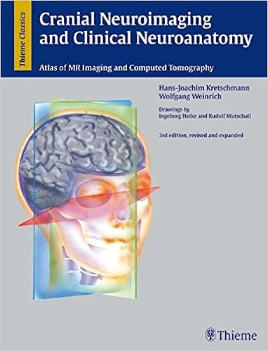
Written through specialists within the box, this fantastically illustrated text/atlas presents the instruments you want to at once visualize and interpret cranial CT and MR pictures. It reports with exacting aspect the traditional anatomic mind constructions pointed out on sagittal, coronal, and axial imaging planes. Use this ebook to make exact and whole neurological checks on the earliest attainable phases - sooner than attaining the sectioning or working table.
This revised and multiplied 3rd version includes approximately six hundred illustrations - such a lot in colour - that offer photo representations of mind buildings, arteries, arterial territories, veins, nerves and neurofunctional platforms. The illustrations depict anatomic buildings in colours of grey just like the best way they're visible in CT and MR images.
Highlights of the 3rd edition:
- Content and illustrations improved by means of greater than 20%
- excessive answer T1 and T2 weighted MR photos
- Improved anatomic terminology for extra actual descriptions of findings
Clinically appropriate, simply readable, and obviously prepared, this well-illustrated e-book is a necessary advent to the sphere for clinical scholars and citizens in neurology, neurosurgery, neuroradiology, and radiology. training experts also will make the most of this functional day by day tool.
Read or Download Cranial Neuroimaging and Clinical Neuroanatomy: Atlas of MR Imaging and Computed Tomography PDF
Similar radiology & nuclear medicine books
Medizinische Physik 3: Medizinische Laserphysik
Die medizinische Physik hat sich in den letzten Jahren zunehmend als interdisziplinäres Gebiet profiliert. Um dem Bedarf nach systematischer Weiterbildung von Physikern, die an medizinischen Einrichtungen tätig sind, gerecht zu werden, wurde das vorliegende Werk geschaffen. Es basiert auf dem Heidelberger Kurs für medizinische Physik.
New advancements akin to sophisticated mixed modality methods and critical technical advances in radiation remedy making plans and supply are facilitating the re-irradiation of formerly uncovered volumes. hence, either palliative and healing methods will be pursued at quite a few disorder websites.
Merrill's Atlas of Radiographic Positions & Radiologic Procedures, Vol 3
Widely known because the best of positioning texts, this highly-regarded, accomplished source good points greater than four hundred projections and ideal full-color illustrations augmented by means of MRI photographs for additional aspect to augment the anatomy and positioning shows. In 3 volumes, it covers initial steps in radiography, radiation safeguard, and terminology, in addition to anatomy and positioning info in separate chapters for every bone crew or organ process.
Esophageal Cancer: Prevention, Diagnosis and Therapy
This publication stories the new development made within the prevention, analysis, and remedy of esophageal melanoma. Epidemiology, molecular biology, pathology, staging, and diagnosis are first mentioned. The radiologic evaluation of esophageal melanoma and the function of endoscopy in analysis, staging, and administration are then defined.
- Cardiac Pacing and ICDS (4th Edition)
- Interventional Oncology (Practical Guides in Interventional Radiology)
- Health Care Reform in Radiology
- Aids to Radiological Differential Diagnosis, 4th Edition
Extra info for Cranial Neuroimaging and Clinical Neuroanatomy: Atlas of MR Imaging and Computed Tomography
Sample text
In MRI the dura mater is demonstrated only after administration of contrast medium. The dura is related to the interhemispheric cistern (Figs. l) and to the transverse cerebral fissure, which is the space between the occipital lobe and the cerebellum (Fig. ll). These spaces are both easily discerned and hence are important guideline structures. Calcifications of the dura mater usually give no MR signal but ossifications may produce a low or, rarely, a high signal. 6 Clinical Value of the New Imaging Techniques The new imaging techniques (CT, CTA, MRI, MRA, PET, SPECT, and ultrasound) have changed our way of thinking and our method of practicing clinical medicine.
The tongue is cut at the level of the hyoid bone. 37 Coronal Slices 1 2 4 5 6 7 8 9 10 11 12 13 14 15 16 Fig. 7d Coronal, T2-weighted MR image. Nonconformity with the sectional plane of Figs. 7a and 7c is shown by the frontal (anterior) horns of the lateral ventricles and the sphenoidal sinus. Superior sagittal sinus Interhemispheric cistern Superior frontal gyrus Middle frontal gyrus Cingulate gyrus Pericallosal artery Genu of corpus callosum Frontal (anterior) horn of lateral ventricle Anterior cerebral artery Inferior frontal gyrus Orbital gyrus Straight gyrus Optic nerve Pole of temporal lobe Inferior nasal concha Atlas 38 1 2 3 4 5 6 7 8 9 10 11 12 13 14 15 16 17 18 19 20 21 22 23 24 25 26 27 28 29 30 31 Superior frontal gyrus Falx cerebri Middle frontal gyrus Cingulate sulcus Cingulate gyrus Inferior frontal gyrus Genu of corpus callosum Frontal (anterior) horn of lateral ventricle Head of caudate nucleus Insula Lateral sulcus (Sylvian fissure) Putamen Superior temporal gyrus Straight gyrus Olfactory tract Optic chiasm Optic nerve Oculomotor nerve Trochlear nerve Middle temporal gyrus Ophthalmic nerve Abducens nerve Sphenoidal sinus Maxillary nerve Nasopharynx Uvula Inferior alveolar nerve Palatine tonsil Lingual nerve Isthmus of fauces Hypoglossal nerve Fig.
The tectal (quadrigeminal) plate lies w i t h i n t h e section and is, therefore, not visible (Fig. 2b). 55 Coronal Slices 1 2 3 4 5 6 7 8 9 10 11 12 13 14 15 16 17 18 19 20 21 22 23 24 25 26 27 28 29 Fig. 12b Coronal, T1-weighted MR image corresponding approximately to t h e sectional plane of Figs. 12a and 12c. Superior sagittal sinus Superior frontal gyrus Precentral gyrus Central sulcus Postcentral gyrus Cingulate gyrus Trunk (body) of corpus callosum Central part (body) of lateral ventricle Supramarginal gyrus Lateral sulcus (Sylvian fissure) Fornix and blood vessel Internal cerebral vein Pulvinar of thalamus Transverse temporal gyrus (of Heschl) Superior temporal gyrus Pineal gland Hippocampus Temporal (inferior) horn of lateral ventricle Middle temporal gyrus Midbrain Parahippocampal gyrus Lateral occipitotemporal gyrus Inferior temporal gyrus Pons Middle cerebellar peduncle Cerebellum Medulla oblongata Spinal cord Vertebral artery 56 1 2 3 4 5 6 7 8 9 10 11 12 13 14 15 16 17 18 19 20 21 22 23 24 25 26 27 28 29 30 31 32 33 Atlas Superior sagittal sinus Paracentral artery Artery of central sulcus Precuneal artery Anterior parietal artery Parietal bone Pericallosal artery Superior thalamostriate (terminal) vein Angular artery Internal cerebral vein Temporal artery of middle cerebral artery Posterior choroidal artery Medial occipital artery Basal vein (of Rosenthal) Lateral occipital artery Superior cerebellar artery Auricle (pinna) Temporal bone Sigmoid sinus Posterior inferior cerebellar artery (PICA) Vertebral artery Occipital bone Mastoid process Atlanto-occipital joint Occipital artery Posterior belly of digastric Vertebral vein Lateral mass of atlas Articular process and arch of axis Articular process and arch of third cervical vertebra Articular process and arch of fourth cervical vertebra Sternocleidomastoid Fifth cervical vertebra Fig.


