By George Simon and W. J. Hamilton (Auth.)
Read or Download X-ray Anatomy PDF
Similar radiology & nuclear medicine books
Medizinische Physik 3: Medizinische Laserphysik
Die medizinische Physik hat sich in den letzten Jahren zunehmend als interdisziplinäres Gebiet profiliert. Um dem Bedarf nach systematischer Weiterbildung von Physikern, die an medizinischen Einrichtungen tätig sind, gerecht zu werden, wurde das vorliegende Werk geschaffen. Es basiert auf dem Heidelberger Kurs für medizinische Physik.
New advancements corresponding to subtle mixed modality methods and important technical advances in radiation remedy making plans and supply are facilitating the re-irradiation of formerly uncovered volumes. accordingly, either palliative and healing ways should be pursued at quite a few sickness websites.
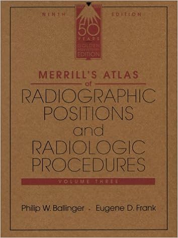
Merrill's Atlas of Radiographic Positions & Radiologic Procedures, Vol 3
Widely known because the top of the line of positioning texts, this highly-regarded, complete source positive aspects greater than four hundred projections and perfect full-color illustrations augmented through MRI photographs for extra element to augment the anatomy and positioning displays. In 3 volumes, it covers initial steps in radiography, radiation defense, and terminology, in addition to anatomy and positioning info in separate chapters for every bone team or organ process.
Esophageal Cancer: Prevention, Diagnosis and Therapy
This e-book experiences the new development made within the prevention, prognosis, and therapy of esophageal melanoma. Epidemiology, molecular biology, pathology, staging, and diagnosis are first mentioned. The radiologic review of esophageal melanoma and the function of endoscopy in analysis, staging, and administration are then defined.
Additional resources for X-ray Anatomy
Example text
Terminal phalanx with rounded end in a normal adult Figure 17. Terminal phalanx with tufted (spade-like) end in a normal adult Figure 18. Second metacarpal of normal adult. Dark lines show points at which the width of the cortex is measured. upper surface towards the anterior end of the second rib {see Figure 214), This is due to the insertion of the second head of the scalenus anterior tendon. The female pelvis tends to be wider than the male {see page 70). There' is minor variation in the length/breadth relationship of the long bones; in some persons they are relatively long and thin, while in others they are shorter and wider.
The ossified acromial end of the clavicle is separated by a wide gap from the acromion, much of which is not yet ossified Figure 53. Radiograph of the shoulder region of a girl aged 9 years, showing the anterior and posterior edges of the epiphyseal line at separate levels. ) Each of the transparent lines of cartilage is bounded above and below by the thin white line of the metaphysis Figure 54. Radiograph of the same girl with the arm slightly flexed. The anterior and posterior edges of the epiphyseal line are seen nearly superimposed.
The x-ray beam is tangential to the glenoid cavity, which now appears as a single white line with the anterior and posterior margins superimposed. The sinous form of the clavicle is recog nizable on account of the rotation of the clavicle that accompanies the movement Figure 42. Radiograph of the same shoulder as in Figure 40, in a shrugged position (posterior view). The acromial end of the clavicle is elevated and this bone is now almost 45 degrees to the horizontal In many subjects an even greater elevation of the scapula may be seen with this movement.


