By Deborah Levine
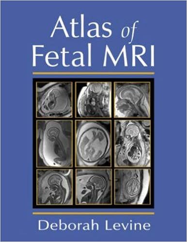
The one textual content to supply in-depth illustrations of the conventional and irregular fetal anatomy on MR imaging, this advisor contains chapters highlighting the state-of-the-science within the imaging of the fetal cranium, face, neck, frightened process, chest, stomach, and musculoskeletal process. Discussing functions on the vanguard of the self-discipline, this reference stories information gleaned from MR examinations of maternal and fetal future health, reports universal quick imaging concepts, info pitfalls regarding fetal MR imaging, and analyzes equipment for bettering picture answer.
Read or Download Atlas of Fetal MRI PDF
Best radiology & nuclear medicine books
Medizinische Physik 3: Medizinische Laserphysik
Die medizinische Physik hat sich in den letzten Jahren zunehmend als interdisziplinäres Gebiet profiliert. Um dem Bedarf nach systematischer Weiterbildung von Physikern, die an medizinischen Einrichtungen tätig sind, gerecht zu werden, wurde das vorliegende Werk geschaffen. Es basiert auf dem Heidelberger Kurs für medizinische Physik.
New advancements corresponding to subtle mixed modality methods and demanding technical advances in radiation therapy making plans and supply are facilitating the re-irradiation of formerly uncovered volumes. subsequently, either palliative and healing ways will be pursued at a number of illness websites.
Merrill's Atlas of Radiographic Positions & Radiologic Procedures, Vol 3
Widely known because the optimum of positioning texts, this highly-regarded, finished source beneficial properties greater than four hundred projections and perfect full-color illustrations augmented through MRI pictures for extra aspect to augment the anatomy and positioning shows. In 3 volumes, it covers initial steps in radiography, radiation defense, and terminology, in addition to anatomy and positioning details in separate chapters for every bone staff or organ method.
Esophageal Cancer: Prevention, Diagnosis and Therapy
This publication studies the hot growth made within the prevention, analysis, and therapy of esophageal melanoma. Epidemiology, molecular biology, pathology, staging, and analysis are first mentioned. The radiologic evaluation of esophageal melanoma and the function of endoscopy in analysis, staging, and administration are then defined.
- The Cell Nucleus. Volume 2
- Human Sectional Anatomy: Pocket Atlas of Body Sections, CT and MRI Images, Third Edition
- Imaging in Oncology (Cancer Treatment and Research)
- Basic Radiology
- Radiation Protection in Medical Imaging and Radiation Oncology (Series in Medical Physics and Biomedical Engineering)
- MRI of the Brain, Head, Neck and Spine: A teaching atlas of clinical applications (Series in Radiology)
Extra info for Atlas of Fetal MRI
Example text
This should not be considered as an abnormal finding, as long as the overall contour and size of the ventricles appears normal, especially during the first two trimesters (Fig. 4). CAVUM OF THE SEPTUM PELLUCIDUM AND CAVUM VERGAE The cavum of the septum pellucidum should always be observed after 20 weeks (Figs. 6). 14 Normal anatomy on coronal T2-weighted image at 38 weeks gestational age. With maturation there has been further reduction in contrast between the gray and white matter and the cerebrospinal fluid spaces are less conspicuous.
Neurosurgery 1996; 39:110– 116. Cardoza JD, Goldstein RB, Filly RA. Exclusion of fetal ventriculomegaly with a single measurement: the width of the lateral ventricular atrium. Radiology 1988; 169:711– 714. Trop I, Levine D. Normal fetal anatomy as visualized with fast magnetic resonance imaging. Top Magn Reson Imaging 2001; 12:3– 17. Levine D, Trop I, Mehta TS et al. MR imaging appearance of fetal cerebral ventricular morphology. Radiology 2002; 223:652– 660. Stazzone MM, Hubbard AM, Bilaniuk LT et al.
9 Mild ventriculomegaly with enlarged cisterna magna and normal cerebellum at 20 weeks gestational age. Sagittal (a), axial (b), and coronal (c) T2-weighted images show mild ventriculomegaly, which is stable at 29 weeks gestational age on sagittal (d) and axial views (e and f ). The cisterna magna is enlarged (arrows); however, the cerebellum appears normal. Note the normal cortical development at 29 weeks. DISORDERS OF DORSAL NEURAL TUBE DEVELOPMENT The CNS develops from the embryonic ectoderm termed the neural plate.


