By M. Chen, T. Pope, D. Ott(eds0
Read or Download Basic Radiology PDF
Similar radiology & nuclear medicine books
Medizinische Physik 3: Medizinische Laserphysik
Die medizinische Physik hat sich in den letzten Jahren zunehmend als interdisziplinäres Gebiet profiliert. Um dem Bedarf nach systematischer Weiterbildung von Physikern, die an medizinischen Einrichtungen tätig sind, gerecht zu werden, wurde das vorliegende Werk geschaffen. Es basiert auf dem Heidelberger Kurs für medizinische Physik.
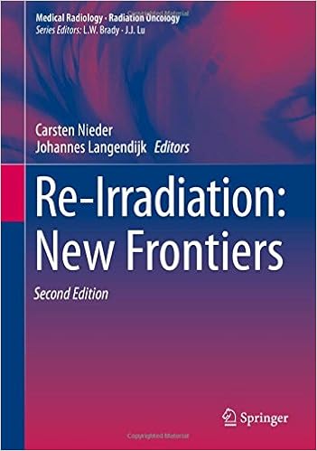
New advancements equivalent to sophisticated mixed modality techniques and important technical advances in radiation remedy making plans and supply are facilitating the re-irradiation of formerly uncovered volumes. for that reason, either palliative and healing methods should be pursued at numerous ailment websites.
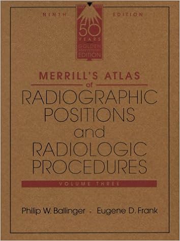
Merrill's Atlas of Radiographic Positions & Radiologic Procedures, Vol 3
Widely known because the top-quality of positioning texts, this highly-regarded, accomplished source positive aspects greater than four hundred projections and perfect full-color illustrations augmented by way of MRI photos for additional element to reinforce the anatomy and positioning displays. In 3 volumes, it covers initial steps in radiography, radiation safeguard, and terminology, in addition to anatomy and positioning info in separate chapters for every bone team or organ procedure.
Esophageal Cancer: Prevention, Diagnosis and Therapy
This booklet stories the hot growth made within the prevention, prognosis, and remedy of esophageal melanoma. Epidemiology, molecular biology, pathology, staging, and analysis are first mentioned. The radiologic overview of esophageal melanoma and the position of endoscopy in analysis, staging, and administration are then defined.
- Cancer Nanotechnology: Principles and Applications in Radiation Oncology (Imaging in Medical Diagnosis and Therapy)
- Radiotherapy Treatment Planning: Linear-Quadratic Radiobiology
- Proton and Charged Particle Radiotherapy
- Ultrasound Teaching Manual: The Basics of Performing and Interpreting Ultrasound Scans
Extra resources for Basic Radiology
Sample text
Exercise 3-3: Pulmonary Vascularity Clinical Histories: Case 3-11. A 28-year-old man examined in the Emergency Department for chest pain and shortness of breath (Fig. 3â 40) Case 3-12. A 65-year-old woman with a 100-packs-a-year history of smoking (Fig. 3â 41) Case 3-13. An acyanotic 22-year-old man with a systolic murmur (Fig. 3â 42) Case 3-14. A 36-year-old man with asthma (Fig. 3â 43) Case 3-15. A 50-year-old woman with acute shortness of breath (Fig. 3â 44) Fig. 3â 40. Fig. 3â 41. Fig. 3â 42.
Fig. 3â 29. Lateral view of patient in Case 3-4 shows filling in of the retrosternal space by the enlarged right ventricle (arrow) and large right and left pulmonary arteries from the pulmonary arterial hypertension (open arrows). 40 41 Discussion: Pericardial effusion and cardiomyopathy have similar appearances on PA chest radiographs (Cases 3-2 and 3-3). This appearance is often referred to as a globular shape or a water-bottle heart. When this appearance is observed, an echocardiogram is the next best imaging test to differentiate between these two entities.
Malposition of the tip. B. pneumothorax. C. perforation. D. catheter coiling. E. catheter thrombosis. 3-27. The complication of CVP catheter placement in Case 3-27 (Fig. 3â 61) is A. malposition of the tip. B. pneumothorax. C. perforation. D. catheter coiling. E. catheter thrombosis. 3-28. The malpositioned catheter in Case 3-28 (Fig. 3â 62) is a(n) A. tracheostomy tube. B. intra-aortic balloon pump. C. Swan-Ganz catheter. 61 62 D. nasogastric tube. E. none of the above. 3-29. Possible complications from the pacemaker shown in Case 3-29 (Fig.


