By Anca-Ligia Grosu, Carsten Nieder
The major aim of this ebook is to supply radiation oncologists with a transparent, updated advisor to tumor delineation and contouring of organs in danger. With this in brain, an in depth assessment of modern advances in imaging for radiation remedy making plans is gifted. Novel ideas for goal quantity delineation are defined, taking into consideration the recommendations in imaging know-how. particular cognizance is paid to the function of the more moderen imaging modalities, akin to positron emission tomography and diffusion and perfusion magnetic resonance imaging. all the most vital tumor entities handled with radiation remedy are coated within the booklet. every one bankruptcy is dedicated to a specific tumor variety and has been written via a famous specialist in that topic.
Read Online or Download Target Volume Definition in Radiation Oncology PDF
Best radiology & nuclear medicine books
Medizinische Physik 3: Medizinische Laserphysik
Die medizinische Physik hat sich in den letzten Jahren zunehmend als interdisziplinäres Gebiet profiliert. Um dem Bedarf nach systematischer Weiterbildung von Physikern, die an medizinischen Einrichtungen tätig sind, gerecht zu werden, wurde das vorliegende Werk geschaffen. Es basiert auf dem Heidelberger Kurs für medizinische Physik.
New advancements akin to sophisticated mixed modality methods and important technical advances in radiation therapy making plans and supply are facilitating the re-irradiation of formerly uncovered volumes. for that reason, either palliative and healing ways might be pursued at a variety of illness websites.
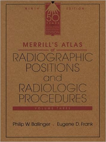
Merrill's Atlas of Radiographic Positions & Radiologic Procedures, Vol 3
Widely known because the optimal of positioning texts, this highly-regarded, finished source gains greater than four hundred projections and ideal full-color illustrations augmented by way of MRI pictures for extra element to reinforce the anatomy and positioning shows. In 3 volumes, it covers initial steps in radiography, radiation security, and terminology, in addition to anatomy and positioning details in separate chapters for every bone workforce or organ approach.
Esophageal Cancer: Prevention, Diagnosis and Therapy
This booklet reports the new development made within the prevention, analysis, and therapy of esophageal melanoma. Epidemiology, molecular biology, pathology, staging, and diagnosis are first mentioned. The radiologic evaluation of esophageal melanoma and the position of endoscopy in analysis, staging, and administration are then defined.
- Intensity-Modulated Radiation Therapy: Clinical Evidence and Techniques
- Diagnostic Imaging: Ultrasound, 1e
- Image-Guided Percutaneous Spine Biopsy
- Simplified Interpretation of ICD Electrograms
- Atlas of FFR-Guided Percutaneous Coronary Interventions
Additional info for Target Volume Definition in Radiation Oncology
Example text
6. 4 Pituitary Adenoma These adenomas are common benign tumours that arise from gland cells of the anterior pituitary lobe. T. Astner et al. 32 Fig. 6 MRI and CT of a patient treated for vestibularis schwannoma by radiosurgery. PTV in pink. 6). Further subtypes are determined according to their ultrastructural differences in electron microscopy. Imaging and target volume definition are different for pituitary micro- or macroadenoma. For radiosurgery of microadenoma adequate imaging is of high priority.
Mandibularis (cranial nerve V3). 5 Canalis caroticus, internal carotid artery. 6 Meatus acusticus externus. 7 Meatus acusticus internus, N. facialis and N. vestibulocochlearis (cranial nerves VII and VIII). 8 Foramen rotundum, N. 2 Tumours of the orbit and their exact anatomical localisation Anatomical structure Extraconal space Surrounding bony structures, periost Fat/conjunctive tissue Localisation Pathology Bony orbital borders Tear system Lacrimal gland Primary bone or chondroid neoplasias, metastasis, infiltration of sinonasal tumours Lymphoma, lipoma, lymphangioma, capillary haemangioma, dermoid/epidermoid, rhabdomyosarcoma Lymphoma, epithelial tumours, sarcoidosis, pseudotumour Intraconal space Muscle tissue, mesenchymal tissue Extraocular muscles Fat/conjunctive tissue Intraconal fat Optic nerve Neural tissue, glial cells Entire route of optic nerve Dura Ocular bulb Retina Choroidea, ciliary body Extraconal tissue Optic sheath Retina, posterior lining of the bulb Choroidea, ciliary body will be described for each tumour entity.
A complete description of these definitions can be found in the ICRU 50 report (ICRU 1993). In addition, the approaches described with respect to dose selection and volume contouring are those currently in practice at the University of Toronto and subject to change. At this time there is a lack of data and consensus as to optimal margins, and the reader should be aware of the limitations in the literature. Case-Based Discussion of Common Spinal Cord Tumors IMST of all three glial lines can occur in the spinal cord.


